Premium  Download Edit
Download the Spinal Cord Facts & Worksheets
Click the button below to get instant access to these worksheets for use in the classroom or at a home.
Download This Worksheet
This download is exclusively for KidsKonnect Premium members!
To download this worksheet, click the button below to signup (it only takes a minute) and you'll be brought right back to this page to start the download!
Sign Me Up
Edit This Worksheet
Editing resources is available exclusively for KidsKonnect Premium members.
To edit this worksheet, click the button below to signup (it only takes a minute) and you'll be brought right back to this page to start editing!
Sign Up
Not ready to purchase a subscription? Click to download the free sample version Download sample
Download This Sample
This sample is exclusively for KidsKonnect members!
To download this worksheet, click the button below to signup for free (it only takes a minute) and you'll be brought right back to this page to start the download!
Sign Me Up
Table of Contents
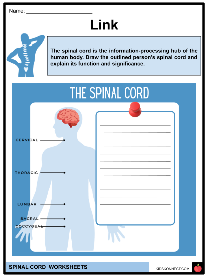
The spinal cord is crucial in connecting the body to the brain. It plays a vital role in relaying information between the brain and the rest of the body. The spinal cord is a long, thin tube of nerve tissue consisting of neurons that extends from the medulla oblongata in the brainstem to the vertebral column of the backbone.
See the fact file below for more information on the Spinal Cord, or you can download our 26-page Spinal Cord worksheet pack to utilize within the classroom or home environment.
Key Facts & Information
Description
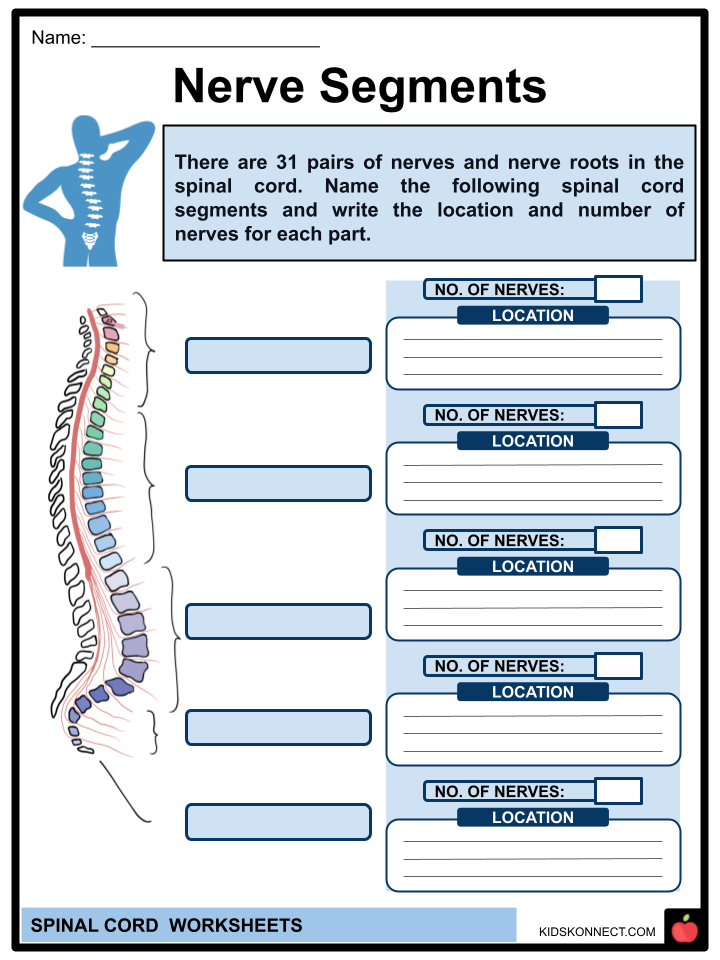
- The spinal cord measures 40 to 50 cm in length and 1 to 1.5 cm in diameter. On each of its sides, two rows of nerve roots emerge. There are 31 spinal nerves, each of which originates from one of these nerve roots.
- The spinal cord is a cylindrical structure of neural tissue formed of white and gray matter that is evenly organized and separated into four regions, cervical, thoracic, lumbar, and sacral, each of which is made up of multiple segments. The spinal nerve is the main conduit for sensory and motor information to and from the brain and the rest of the body. Each spinal cord segment innervates a dermatome.
Function
- The spinal cord manages body functions and movements. Movements are governed by the transmission of signals from the brain to the rest of the body. They also control autonomic or involuntary activities such as respiratory rate, heart rate, and bowel and bladder function.
- It sends sensory data to the brain. Signals from different body regions assist the brain in registering and processing sensations such as pressure and pain. It also manages your reflexes. The spinal cord controls certain reflexes (involuntary movements) independently of the brain. One reflex governed by the spinal cord is the patellar reflex, or the forced leg movement when someone taps the shin in a specific spot.
Anatomy
External: Spinal Cord Nerves
- There are 31 pairs of nerves and nerve roots in the spinal cord. Beginning at the neck and descending to the buttocks (back end), the following portions include:
- Cervical (nerves starting in your neck and mainly running to your face and head): The cervical region of the spine consists of eight segments; thus, it has eight cervical nerves (C1 to C8/T1). The cervical vertebrae allow for head rotation, tilting, and nodding. A lordotic curve is the inward C-shape formed by the cervical spine.
- Thoracic (nerves in your upper body that extend to your chest, upper back, and abdomen): There are 12 thoracic nerves in the trunk or thoracic region of the spine (T1 to T12). The thoracic spine is where your ribs attach. The kyphotic curve is a little outward curvature of this portion of the spine that forms a backward C-shape.
- Lumbar (nerves in the low back that run to your legs and feet): The lumbar region of the spine consists of five segments, so it has five lumbar nerves (L1 through L5) that supports the body’s weight. The lumbar spine provides support for the higher portions of the spine. It’s where the pelvis is attached, taking the brunt of your body’s weight and the strain of lifting and carrying. The lumbar spine is the site of numerous back issues. The lumbar spine curves inward to form a C-shaped lordosis.
- Sacral (nerves in the low back extending into the pelvis): There are five sacral segments and thus five sacral nerves (S1 to S5).
- Coccygeal (tailbone): This little portion of bone near the spine’s base comprises four joined vertebrae. The coccyx is where the ligaments and muscles of the pelvic floor meet. It consists of one coccygeal segment.
Internal: Structure of a Spinal Cord
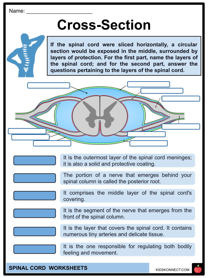
- The spinal cord, like the brain, is coated with tissue layers known as meninges. These include the dura mater, the arachnoid mater, and the pia mater. The medical term for these layers is meninges.
- Dura mater: The dura mater is the outermost layer of the meninges of the spinal cord. It is a solid and protective coating.
- Epidural space: The epidural space is the area containing fat and small arteries between the dura mater and the spinal wall. In this area, cerebrospinal fluid covers the spinal cord and brain stem at the periphery of the dural sac.
- Arachnoid mater: This comprises the middle layer of the spinal cord’s covering. It is a fluid-filled weblike structure that cushions the brain.
- Subarachnoid space: This area is found between the arachnoid mater and the pia mater. Cerebrospinal fluid (CSF) resides in this area. CSF samples are often taken to check for infections like meningitis, which can be fatal if left untreated.
- Pia mater: This is the layer that covers the spinal cord. It contains numerous tiny arteries and is made up of delicate connective tissue.
- A horizontal “slice” of the spinal cord would reveal a circular portion surrounded by protective layers in the center. From this circular region, extend nerve projections. This section includes the following:
- Gray matter: The dark, butterfly-shaped area of the spinal cord that contains the bodies of the nerve cells.
- White matter: This is made up of myelinated cells and surrounds the gray matter, allowing for faster nerve communication. Myelin coats nerve cells in a gray matter less densely than in a white case.
- Posterior root: The portion of a nerve that emerges from behind the spinal column is called the posterior root. In a cross-section of the spinal cord, the upper wings of the “butterfly” of gray matter extend toward the vertebrae. The bottom branches are positioned toward the body’s front and internal organs.
- Anterior root: The segment of the nerve that emerges from the front of the spinal column is called the anterior root.
- Spinal ganglion: The sensory neurons are grouped in the spinal ganglion, a collection of nerve cells.
- Spinal nerve: A spinal nerve results from the union of the spinal column’s posterior and anterior roots. There are 31 spinal nerve pairs. These regulate both bodily feeling and movement.
Spinal Cord Injuries
- Any injury to the spinal cord or spinal nerves causes chronic and lifelong damage to the spinal cord, impairing its ability to operate normally without replacement. It frequently causes permanent alterations in the body’s strength, posture, and reflexes. The extent and location of an injury affect how much control a person has over their limbs once injured.
- Injuries to the spinal cord affect not just the spinal neurons and vertebral column but also other muscles and essential organs. Two types of paralysis can result from an injury:
- Tetraplegic – Paralysis characterized by entire or partial loss of function in all four limbs and the trunk.
- Paraplegic – The only difference between paraplegia and tetraplegia is that paraplegia does not affect the arms.
Spinal Cord Worksheets
This is a fantastic bundle that includes everything you need to know about the Spinal Cord across 26 in-depth pages. These are ready-to-use worksheets that are perfect for teaching kids about the Spinal Cord, a long, thin tube of nerve tissue consisting of neurons that extends from the medulla oblongata in the brainstem to the vertebral column of the backbone.
Complete List of Included Worksheets
Below is a list of all the worksheets included in this document.
- Spinal Cord Facts
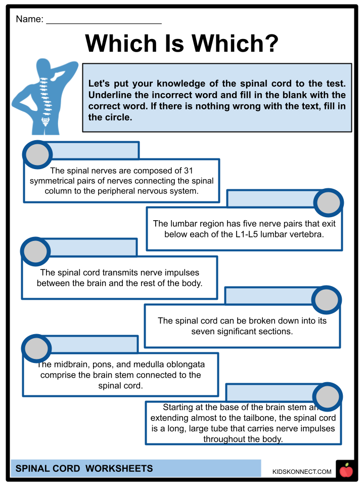
- Which Is Which?
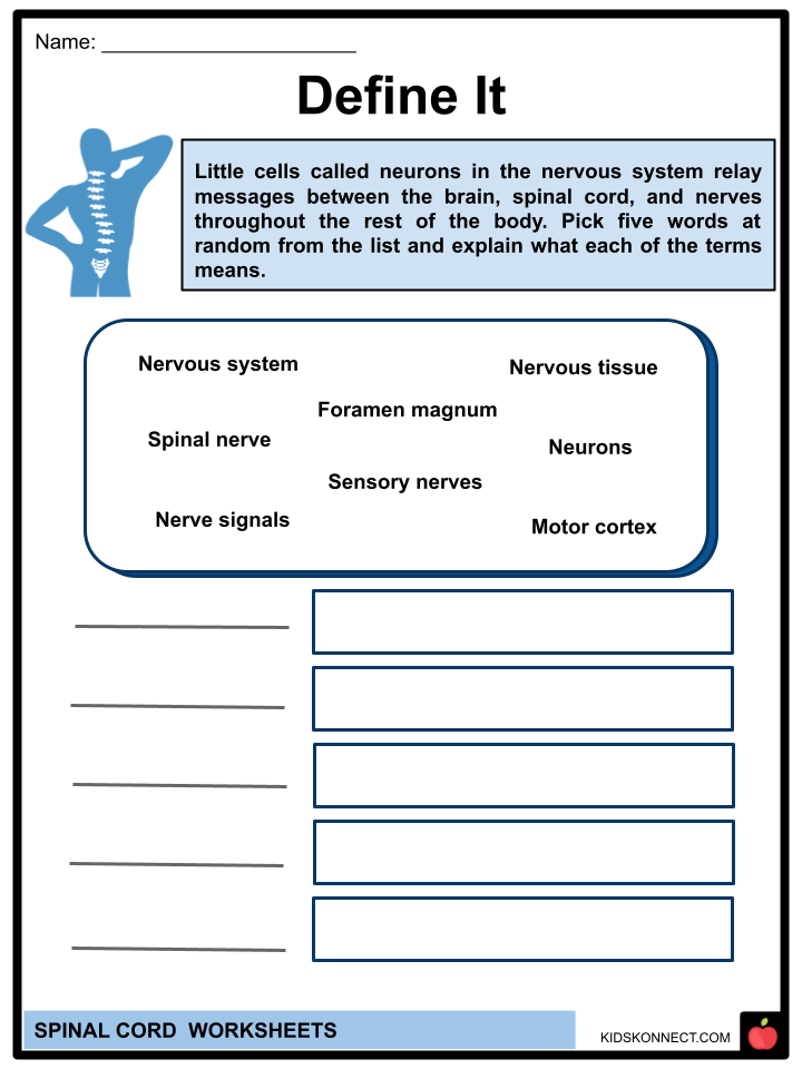
- Define It
- Nerve Segments
- Describe It
- Cross-Section
- Link
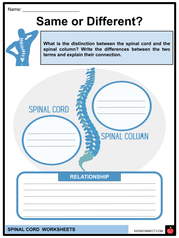
- Same or Different?
- Trivia Facts
- Spinal Injury
- Healthy Practice
Link/cite this page
If you reference any of the content on this page on your own website, please use the code below to cite this page as the original source.
Link will appear as Spinal Cord Facts & Worksheets: https://kidskonnect.com - KidsKonnect, November 10, 2022
Use With Any Curriculum
These worksheets have been specifically designed for use with any international curriculum. You can use these worksheets as-is, or edit them using Google Slides to make them more specific to your own student ability levels and curriculum standards.


