Premium  Download Edit
Download the Heart Facts & Worksheets
Click the button below to get instant access to these worksheets for use in the classroom or at a home.
Download This Worksheet
This download is exclusively for KidsKonnect Premium members!
To download this worksheet, click the button below to signup (it only takes a minute) and you'll be brought right back to this page to start the download!
Sign Me Up
Edit This Worksheet
Editing resources is available exclusively for KidsKonnect Premium members.
To edit this worksheet, click the button below to signup (it only takes a minute) and you'll be brought right back to this page to start editing!
Sign Up
Not ready to purchase a subscription? Click to download the free sample version Download sample
Download This Sample
This sample is exclusively for KidsKonnect members!
To download this worksheet, click the button below to signup for free (it only takes a minute) and you'll be brought right back to this page to start the download!
Sign Me Up
Table of Contents
Make a fist with your hands then, squeeze it about five times. That motion is similar to what your heart does to pump blood throughout the body. Without the heart, all our body parts will not be able to function.
See the fact file below for more information on Heart or alternatively, you can download our 27-page Heart worksheet pack to utilize within the classroom or home environment.
Key Facts & Information
WHAT IS THE HEART?
- The heart is the central organ of the circulatory system. It is a muscular organ responsible for pumping blood throughout the body.
- As it pumps blood, allows the transportation of nutrients and gases to and from the different parts of the body.
- Hearts differ for each organism but they basically have the same function. Humans, as well as other mammals and birds, have four-chambered hearts. Fish have two chambers while reptiles have three.
- In this fact file, we will be focusing on the human heart.
THE HUMAN HEART
- Place your right hand on your left chest. The location of your palm is the approximate location of your heart.
- It is located in between your lungs, resting slightly on the left. This location is also the reason why our right lung is slightly bigger than the left. The heart, and the lungs, are protected by our rib cage.
- Since it is surrounded by other organs, such as the lungs that are continuously contracting and relaxing, it needs protection. The pericardium that covers it provides this muscular organ with a frictionless environment.
THE HEART WALL AND ITS LAYERS
- The heart wall is composed of three layers: epicardium, myocardium, and endocardium. The epicardium is found below the pericardium. Just like the pericardium, the epicardium functions to protect the heart from injury and even ensures that cardiac cells develop properly.
- The myocardium is the layer of muscle cells that make up the walls of the heart. This is found between the epicardium and the endocardium. This layer makes up the bulk of the heart and is responsible for heart contractions.
- Lastly, found under the myocardium is the innermost layer, the endocardium. The endocardium is a thin and smooth membrane that gives the inner surface of the heart its glistening appearance. Aside from this, the endocardium aids in the formation of the valves that prevent the backflow of blood.
THE CHAMBERS OF THE HEART
- Biology tells us that the human heart, as mentioned, is made up of four chambers. These four chambers are the left atrium, left ventricle, right atrium, and right ventricle. The right chambers make up the right part, while the left ones make up the left part.
- The atria (plural for atrium) are the receiving chambers. Meanwhile, the ventricles are the pumping chambers. Due to this difference in function, the ventricles have thicker walls compared to the atria.
- The right part is responsible for receiving and pumping deoxygenated blood, carbon-dioxide containing blood. On the other hand, the left part is responsible for receiving and pumping oxygenated blood, oxygen-containing blood.
VALVES
- Valves are structures found in between the chambers and the blood vessels connected to the heart which has four valves. These Valves ensure that blood will flow from one direction only.
- In between the right atrium and right ventricle is the tricuspid valve. This prevents the backflow of blood from the right ventricle to the right atrium.
- Another is the pulmonary valve, found between the right ventricle and the pulmonary artery. The pulmonary artery connects the right ventricle to the lungs. Thus, this valve prevents backflow to the right ventricle.
- The third valve is called the mitral valve, sometimes the bicuspid. This is located between the left atrium and left ventricle. This prevents backflow to the left ventricle.
- Last but not the least, is the aortic semilunar valve. This valve is located between the left ventricle and the aorta. It prevents the backflow of blood from the aorta to the left ventricle.
- Fun fact, the sound that the doctors are listening to through their stethoscopes is made by the opening and closing of the valves. If you have a stethoscope at home or at school, you can listen to this sound. Simply watch out for the “lub-dub” sound.
- The “lub” sound is made when the tricuspid and mitral valves close. The “dub” sound is made when the aortic and pulmonary valves close.
- These sounds signify that it is working properly. Remember, it is very important that our blood is flowing in one direction only.
THE FLOW OF BLOOD THROUGH THE HEART
- Now that you have learned about its structure, it is time for you to learn how blood flows through the heart and how it will be distributed throughout the body.
- The heart and the different blood vessels connected to it allow blood to be transported and distributed to and from the body.
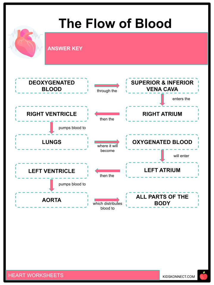
- The blood will drop off oxygen to cells and tissues and will pick up carbon dioxide. This deoxygenated blood will be drained in the right atrium via the superior and inferior vena cava.
- The right atrium will then drain the deoxygenated blood to the right ventricle. The right ventricle will then pump the blood to the lungs for oxygenation via the pulmonary artery. As this happens, the tricuspid valve must be closed to prevent backflow to the right atrium and the pulmonary valve must be open to allow entry to the lungs.
- Upon entry of deoxygenated blood into the lungs, the pulmonary valve must remain closed.
- After getting oxygen from the lungs, the blood, now oxygenated, will return to the heart via the pulmonary vein. The pulmonary vein will drain the oxygenated blood to the left atrium.
- The left atrium will then drain the oxygenated blood to the left ventricle. The left ventricle will then pump the blood to the aorta. As this happens the mitral valve must be closed while the aortic semilunar valve must be open.
- As the oxygenated blood travels to the aorta and to the different parts of the body, the aortic semilunar valve must be closed to prevent blood from going back to the left ventricle.
- The aorta, which is the largest artery, will distribute oxygenated blood to all parts of the body.
- The oxygenated blood will drop off oxygen to cells and tissues and will pick up carbon dioxide. The whole process will repeat as long as the heart is functioning and as long as we are alive.
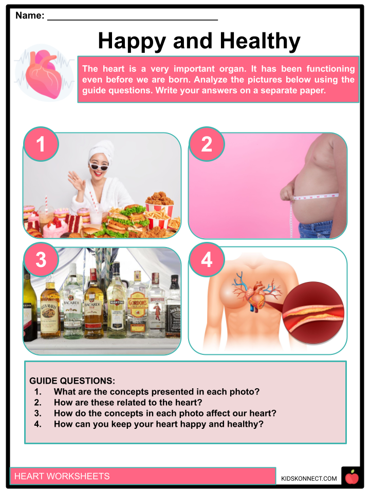
KEEPING YOUR HEART HEALTHY
- The heart is a very important organ, it has been pumping blood even before we were born and will keep on doing so as long as we are living. Thus, it is very important to keep it healthy.
- Although most of us are born with a healthy heart, our lifestyle can affect its health and functioning.
- Physical fitness is very important not just for the muscles that allow us to move but also for the heart. The heart is responsible for replenishing our cells with oxygen and nutrients, and for picking up waste materials.
- Activities that allow our bodies to consume more oxygen than needed will exercise our hearts. It will increase the efficacy of our hearts. So, make sure to participate in activities such as running, jumping, and even dancing.
- Another thing that we can do is by avoiding foods that are fatty. Fat crystals can build up in our blood vessels. This fat buildup will prevent the proper flow of blood, and may even clog our blood vessels. If this happens, certain parts of our body will not be able to receive oxygen and other nutrients, and also won’t be able to remove waste such as carbon dioxide.
- There is a lot more that we can do to keep our heart healthy and happy. Reading articles and listening to health professionals can help us maintain the health of our hearts.
Heart Worksheets
This is a fantastic bundle that includes everything you need to know about Heart across 27 in-depth pages. These are ready-to-use worksheets that are perfect for teaching about the Heart which is the central organ of the circulatory system.
Complete List of Included Worksheets
Below is a list of all the worksheets included in this document.
- Heart Fact File
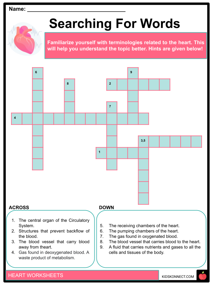
- Searching For Words
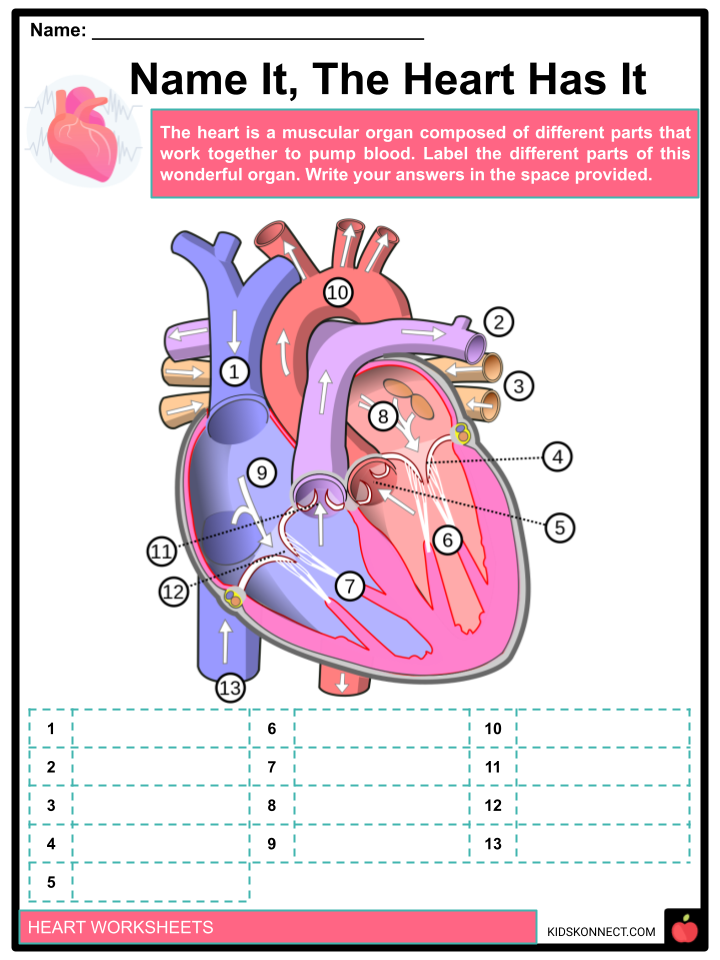
- Name It, The Heart Has It
- The Flow of Blood
- Fact or Bluff
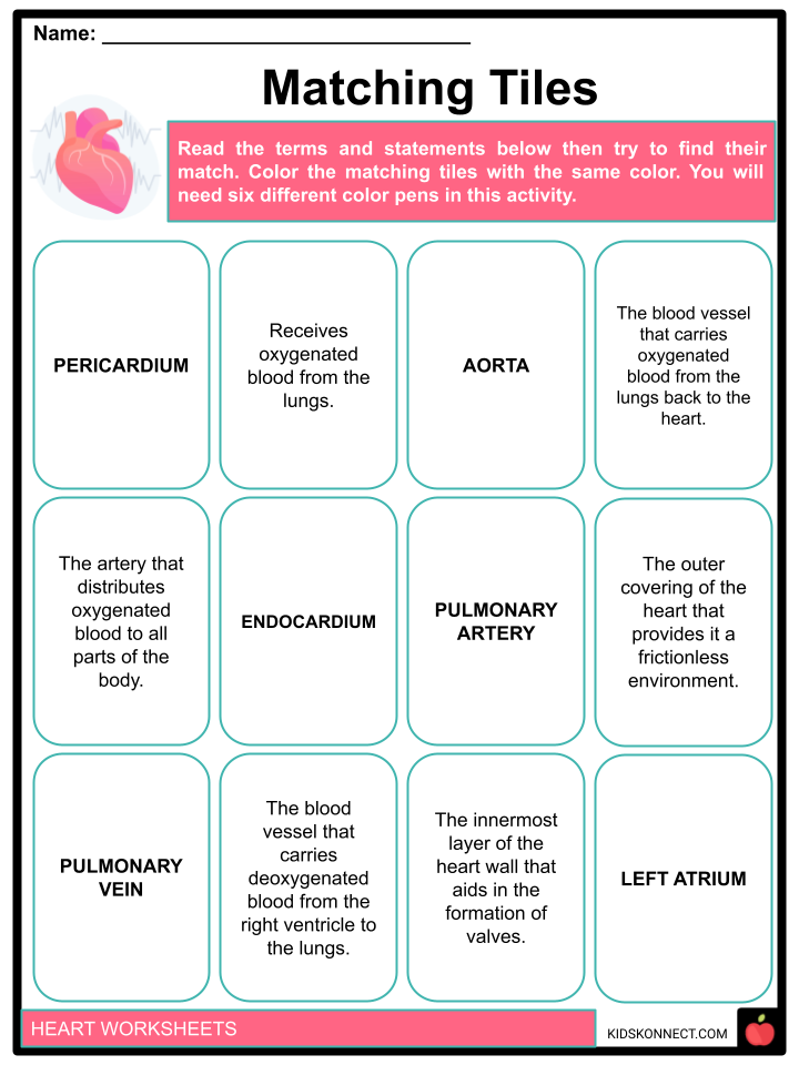
- Matching Tiles
- Keep A Pulse
- Happy and Healthy
- Why It Matters
- Cheers To A Healthy Lifestyle!
- To The Heart
Frequently Asked Questions
What are the four parts of the heart?
The heart has four chambers and they are: the left atrium, left ventricle, right atrium, and right ventricle.
How does the heart function?
It has different blood vessels connected to it that allow blood to be distributed to and from the body with every pump. Blood carries oxygen and other nutrients to our cells and tissues and carries away carbon dioxide and other waste.
What is the size of our heart?
Generally, an adult’s heart is the size of two fists, while a child’s heart is about the size of one clenched fist.
Link/cite this page
If you reference any of the content on this page on your own website, please use the code below to cite this page as the original source.
Link will appear as Heart Facts & Worksheets: https://kidskonnect.com - KidsKonnect, July 20, 2022
Use With Any Curriculum
These worksheets have been specifically designed for use with any international curriculum. You can use these worksheets as-is, or edit them using Google Slides to make them more specific to your own student ability levels and curriculum standards.


