Premium  Download Edit
Download the Neurons Facts & Worksheets
Click the button below to get instant access to these worksheets for use in the classroom or at a home.
Download This Worksheet
This download is exclusively for KidsKonnect Premium members!
To download this worksheet, click the button below to signup (it only takes a minute) and you'll be brought right back to this page to start the download!
Sign Me Up
Edit This Worksheet
Editing resources is available exclusively for KidsKonnect Premium members.
To edit this worksheet, click the button below to signup (it only takes a minute) and you'll be brought right back to this page to start editing!
Sign Up
Not ready to purchase a subscription? Click to download the free sample version Download sample
Download This Sample
This sample is exclusively for KidsKonnect members!
To download this worksheet, click the button below to signup for free (it only takes a minute) and you'll be brought right back to this page to start the download!
Sign Me Up
Table of Contents
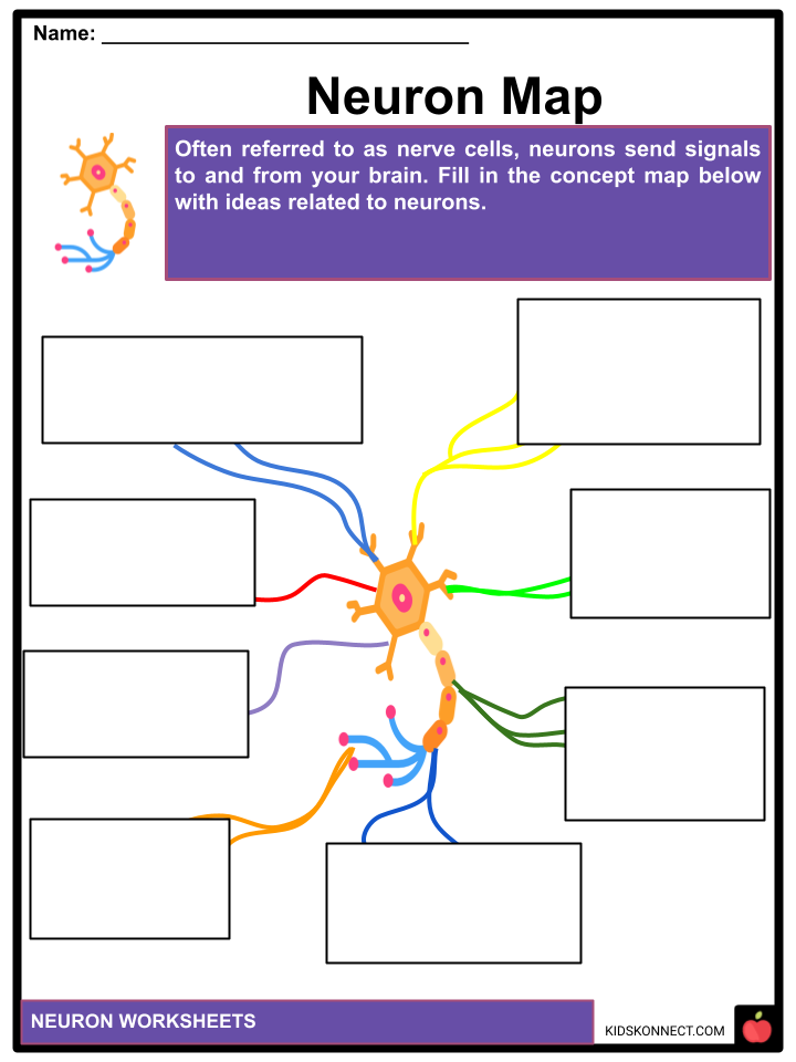
Often referred to as nerve cells, neurons send signals to and from your brain. Neurons are morphologically and functionally unique from other cell types yet have many traits in common with them. The electrically excitable cell known as a neuron shoots electric impulses known as action potentials throughout a neural network.
See the fact file below for more information on Neurons, or you can download our 32-page Neurons worksheet pack to utilize within the classroom or home environment.
Key Facts & Information
HISTORY
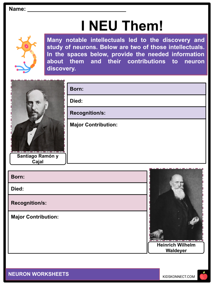
- The Spanish anatomist Santiago Ramon y Cajal’s research in the late 19th century helped establish the neuron as the basic functioning unit of the nervous system. Ramon y Cajal enhanced a technique Camillo Golgi had created for silver staining to reveal the structure of individual neurons. The “double impregnation” method, used in the modified procedure, is still in use.
- The word “neuron” was used by German anatomist Heinrich Wilhelm Waldeyer in 1891 to refer to the anatomical and physiological unit of the nervous system in his examination of the neuron concept. The underlying assumption that neurons are the primary anatomical and functional elements of the nervous system is known as the “neuron doctrine.” Santiago Ramon y Cajal proposed the idea in the late 19th century. It advocated that neurons are separate cells that function as metabolically unique entities.
- The doctrine has improved as a result of recent findings. For instance, non-neuronal glial cells are crucial to the processing of information. Additionally, electrical synapses—direct, cytoplasmic connections between neurons—are more frequent than previously believed.
- Despite being frequently referred to as the “fundamental units” of the brain, neurons carry out internal calculations. In models where neurons are treated as a basic unit, the intricacy of how neurons integrate input inside dendrites is overlooked. Dendritic branches may be seen as spatial compartments, each of which has a unique activity connected to the others owing to passive membrane characteristics but may also alter based on synaptic input.
NERVOUS SYSTEM & NEURONS IN THE BRAIN
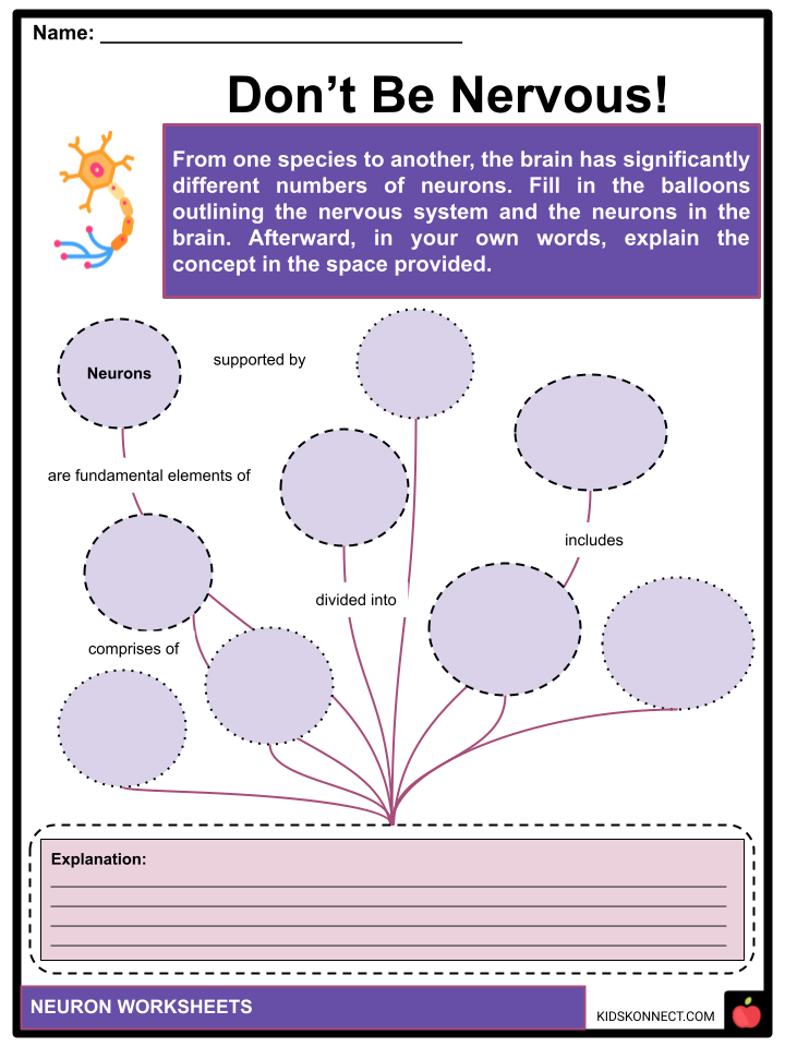
- The fundamental elements of the nervous system are neurons, which are supported structurally and metabolically by glial cells. The peripheral nervous system, which includes the autonomic and somatic nervous systems, and the central nervous system, which comprises the brain and spinal cord, comprise the nervous system.
- From one species to another, the brain has significantly different numbers of neurons. The human cerebral cortex and cerebellum are each thought to have 10–20 billion and 55–70 billion neurons, respectively. The nematode worm Caenorhabditis elegans, in comparison, has just 302 neurons, making it an excellent model organism since all of its neurons have been mapped by scientists.
- Scientists may examine processes happening in more sophisticated creatures in much simpler experimental setups because many characteristics of neurons, including the kind of neurotransmitters employed and the makeup of ion channels, are conserved across species.
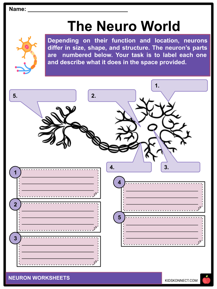
ANATOMY
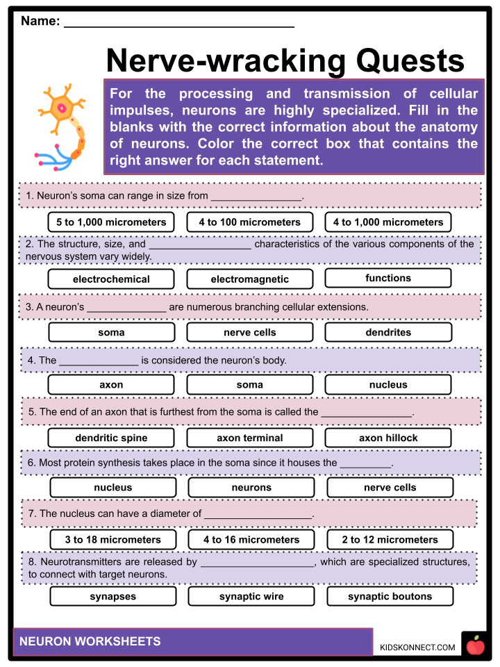
- For the processing and transmission of cellular impulses, neurons are extremely specialized. The structure, size, and electrochemical characteristics of the various components of the nervous system vary widely because of the range of duties they carry out. For instance, a neuron’s soma can range from 4 to 100 micrometers.
- The cell body, or soma, is the central portion of the neuron. The cell body preserves the structural integrity of the neuron, houses the genetic material, and supplies energy to power operations. A neuron’s soma has a nucleus and specialized organelles, much like other cell bodies do. It is protected and given access to its local environment by a membrane surrounding it.
- A neuron’s dendrites are numerous branching cellular extensions. The term “dendritic tree” is used metaphorically to describe this general form and structure. Most of the input to the neuron comes from here via the dendritic spine.
- The soma’s diameter can be tens, hundreds, or even thousands of times less than the diameter of the axon, which is a smaller, cable-like projection.
- In addition to returning some information to the soma, the axon primarily conducts nerve signals away from it. Although many neurons only have one axon, this axon can—and frequently does—go through substantial branching, allowing it to communicate with various target cells.
- Although the axon and axon hillock often play a role in information outflow, other neurons can also send input to this area. The end of the axon that is furthest from the soma is called the axon terminal, and it has sinuses.
- Neurotransmitters are released by synaptic boutons, specialized structures, to connect with target neurons. A neuron may have en passant boutons along the length of the axon in addition to synaptic boutons at the axon terminal.
TYPES OF NEURONS
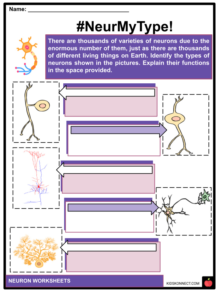
- The structure, function, and genetic makeup of neurons differ. There are thousands of varieties of neurons due to their enormous number, just as there are thousands of different living things on Earth. There are five main types of neurons, however. Each incorporates various components of the fundamental neuron form.
- Unipolar nerve cells – These neurons typically only exist in invertebrate animals and contain a single axon. Their dendrites are picking up sensory data, perhaps right from the stimuli. Unipolar neurons’ cell bodies are always located in the ganglia.
- Bipolar cells – Two extensions protrude from the cell body of bipolar neurons. The axon is located at the tip of one side, while the dendrites are situated on the other. The retina of the eye is where these neurons are most frequently located.
- However, they are also present in areas of the neurological system that support the nose and ears.
- Pyramidal nerve cells – These neurons don’t just have one dendrite; they have numerous, forming a pyramidal structure. The cortex is where you’ll usually find these giant neuron cells. The area of the brain that controls conscious cognition is called the cortex.
- Multipolar nerve cells – These neurons have symmetrical dendrites that branch off a single axon. They are the most prevalent kind of neuron in the central nervous system.
- Purkinje nerve cells – Multiple dendrites branch out from the cell body of Purkinje neurons. Because they produce neurotransmitters that prevent other neurons from firing, these neurons are inhibitory.
- Depending on their intended use, scientists classify neurons into three main categories: sensory, motor, and interneurons.
- Sensory neurons Physical and chemical signals from your surroundings activate sensory neurons. Physical inputs include touch, heat, light, and sound. Chemical inputs are used in smell and taste. For instance, the soles of your feet are activated when you tread on hot sand. Your brain receives a signal from those neurons alerting you to the heat.
Motor neurons
- Movement, both voluntary and involuntary, is regulated by motor neurons. These neurons communicate between the brain and spinal cord and the body’s muscles, glands, and organs.
- Lower and upper motor neurons are two different kinds. The spinal cord sends messages to the smooth and skeletal muscles through lower motor neurons. Between your brain and spinal cord, upper motor neurons convey impulses. Lower motor neurons in your spinal cord, for instance, signal the smooth muscles of your esophagus, stomach, and intestines when you eat. When these muscles flex, food may pass through your digestive system.
- Interneurons You can find interneurons, and neuronal bridges, in your brain and spinal cord. The majority of neurons are of this kind. Motor neurons and other interneurons transmit signals from sensory neurons and other interneurons. They frequently create intricate circuits that facilitate your response to outside inputs.
- For instance, sensory neurons in your fingertips may alert interneurons in your spinal cord when you contact something pointy like a cactus. You can move your hand aside by sending the signal to the motor neurons in your hand through specific interneurons. You feel pain when other interneurons in your brain send a message to the pain center.
CLASSIFICATION
- Neurons can be categorized based on their morphology and function and vary in size and form. The two types of neurons identified by the anatomist Camillo Golgi are type I, which has long axons utilized to transmit impulses over great distances, and type II, which has small axons sometimes mistaken for dendrites.
- The soma’s position allows for further classification of type I cells. The soma, the cell body of type I neurons, and the long, thin axon wrapped by the myelin sheath are their main structural components. Spinal motor neurons represent type I neurons.
- Receiving messages from neighboring neurons, the dendritic tree encircles the cell body. The synaptic cleft, which separates the terminals from the dendrites of the next neuron, is where the branching axon terminals at the end of the axon release neurotransmitters.
FUNCTIONS OF NEURONS
- A neuron’s critical functions are as follows:
- Chemical Synapse – The action potential in chemical synapses impacts other neurons via a space between two neurons identified as the synapse. The action potential is transported up the axon to a postsynaptic terminal, where it begins the release of neurotransmitters, which are chemical messengers. These neurotransmitters stimulate postsynaptic neurons, which create their action potentials.
- Electric Synapse – An electrical synapse is a gap junction joining two neurons. Ion channels in the gaps aid in immediately transferring a positive electrical signal. These occur at a much higher rate than chemical synapses.
CONNECTIVITY
- Synapses are points where the axon terminal of one neuron connects to the dendrite, soma, or, less frequently, the axon of another neuron. Other neurons in the supraoptic nucleus have just one or two dendrites, each of which gets thousands of synapses. For instance, Purkinje cells in the cerebellum link with tens of thousands of other cells through their over 1000 dendritic branches.
- The activity of the target cell can be increased or decreased by synapses depending on whether they are excitatory or inhibitory. Electrical synapses are direct contacts between cells that are electrically conductive and are another method used by specific neurons to communicate.
- Action potentials activate voltage-gated calcium channels at the axon terminal, allowing calcium ions to enter the terminal. The postsynaptic neuron’s receptors are activated by the neurotransmitter as it diffuses across the synaptic cleft.
- Axon terminals with high levels of cytosolic calcium are stimulated to take up calcium, which in turn stimulates mitochondrial energy metabolism to create ATP to enable ongoing neurotransmission.
- Approximately 8.6 × 10¹⁰ (eighty-six billion) neurons comprise the human brain. Each neuron has 7,000 synaptic connections on average. A three-year-old’s brain is thought to have ten synapses (1 quadrillion), on average. With age, this number decreases until maturity, when it stabilizes. There are several estimates for an adult, ranging from 100 to 500 trillion synapses or 1014 to 5×10¹⁴.
NEUROLOGICAL DISORDERS
- Alzheimer’s disease (AD), usually referred to as Alzheimer’s, is a neurodegenerative condition marked by increasing cognitive decline, decreased daily living activities, and behavioral abnormalities or neuropsychiatric symptoms.
- Parkinson’s disease (PD), usually known as Parkinson’s, is a central nervous system degenerative condition that frequently affects speech and movement function. The category of diseases known as movement disorders includes Parkinson’s disease.
- In its advanced phases, Charcot-Marie-Tooth disease (CMT) is a heterogeneous genetic condition of the nerves (neuropathy) that mainly affects the feet and legs but can also affect the hands and arms, causing loss of muscle tissue and touch sensitivity. One of the most prevalent hereditary neurological illnesses, this condition, now incurable, affects 36 in every 100,000 people.
- A neuromuscular condition called myasthenia gravis causes intermittent muscle weakness and tiredness during routine activity. The most common cause of deficiency is circulating antibodies that block acetylcholine receptors at the postsynaptic neuromuscular junction, preventing the neurotransmitter acetylcholine stimulatory effects.
Neuron Worksheets
This fantastic bundle includes everything you need to know about Neurons across 32 in-depth pages. These ready-to-use worksheets are perfect for teaching kids about Neurons. Neurons are morphologically and functionally unique from other cell types yet have many traits in common with them.
Complete List of Included Worksheets
Below is a list of all the worksheets included in this document.
- Neuron Facts
- I NEU Them!
- Neuron Map
- The Neuro World
- Don’t Be Nervous!
- #NeurMyType!
- Nerve-wracking Quests
- Battle of the Nerves!
- NEU Story!
- Oh SyNAPSe!
- Brain Campaign
Frequently Asked Questions
What is a neuron?
A neuron is a specialized cell in the nervous system responsible for transmitting electrical and chemical signals. It is the basic building block of the brain, spinal cord, and the entire nervous system.
What are the main parts of a neuron?
- Neurons consist of three main parts: the cell body (soma), dendrites, and an axon. The cell body contains the nucleus and other essential cellular components, dendrites receive signals from other neurons, and the axon transmits signals away from the cell body to other neurons or target cells.
How do neurons communicate with each other?
Neurons communicate through synapses. When an electrical signal, known as an action potential, reaches the end of an axon, it triggers the release of neurotransmitters. These neurotransmitters cross the synapse, a tiny gap between neurons, and bind to receptors on the dendrites or cell body of the neighboring neuron. This process allows the transmission of information from one neuron to another.
What is the role of myelin in neuron function?
Myelin is a fatty substance that wraps around the axon of certain neurons, forming a protective sheath. It acts as an insulator, speeding up the transmission of electrical signals along the axon. Myelinated neurons conduct signals more efficiently than unmyelinated ones, allowing for faster communication within the nervous system.
How does the firing of a neuron affect its target cells?
When a neuron fires, it generates an electrical impulse called an action potential. This action potential travels down the axon to the synapse, where it triggers the release of neurotransmitters. These neurotransmitters then bind to receptors on the target cells, such as other neurons or muscle cells. Depending on the type of neurotransmitter and the receptors involved, the firing of a neuron can excite or inhibit the activity of the target cell, influencing various physiological and cognitive processes.
Link/cite this page
If you reference any of the content on this page on your own website, please use the code below to cite this page as the original source.
Link will appear as Neurons Facts & Worksheets: https://kidskonnect.com - KidsKonnect, August 19, 2023
Use With Any Curriculum
These worksheets have been specifically designed for use with any international curriculum. You can use these worksheets as-is, or edit them using Google Slides to make them more specific to your own student ability levels and curriculum standards.


