Premium
 Download
Edit
Download
Edit
Download the Trachea Facts & Worksheets
Click the button below to get instant access to these worksheets for use in the classroom or at a home.
Download This Worksheet
This download is exclusively for KidsKonnect Premium members!
To download this worksheet, click the button below to signup (it only takes a minute) and you'll be brought right back to this page to start the download!
Sign Me Up
Edit This Worksheet
Editing resources is available exclusively for KidsKonnect Premium members.
To edit this worksheet, click the button below to signup (it only takes a minute) and you'll be brought right back to this page to start editing!
Sign Up
Not ready to purchase a subscription? Click to download the free sample version Download sample
Download This Sample
This sample is exclusively for KidsKonnect members!
To download this worksheet, click the button below to signup for free (it only takes a minute) and you'll be brought right back to this page to start the download!
Sign Me Up
Table of Contents
The trachea, often known as the windpipe, is a respiratory organ with a diameter of less than an inch and a length of around 4 inches. The trachea allows the passage of air from the larynx to the lungs.
See the fact file below for more information on the Trachea, or you can download our 27-page Trachea worksheet pack to utilize within the classroom or home environment.
Key Facts & Information
STRUCTURE
- The trachea is a fibrocartilaginous respiratory organ with a D-shaped form. Anterolaterally, it comprises 16–20 tracheal cartilages and, laterally, a fibromuscular wall. Hyaline cartilage makes up the tracheal cartilage, joined together by fibroelastic tissue. When pressure fluctuations are associated with air ventilation, they support the trachea and maintain it open.
- The trachealis muscle shapes the trachea’s posterior wall, giving the cartilage the appearance of incomplete C-shaped rings. The trachea’s wall structure gives it enough flexibility and elasticity to allow for the temporary extension of the esophagus during swallowing.
- There are four histological layers in the trachea. The uppermost layer is the mucosa, which is lined by pseudostratified ciliated columnar epithelium. The submucosa is the second histological layer.
- It comprises connective tissue that houses lymphatics, smooth muscle, arteries, nerves, and mucus glands. The cartilaginous rings and abutting smooth muscle make up the third layer, which is known as the muscular cartilaginous layer. The fibroelastic adventitia provides the outermost layer.
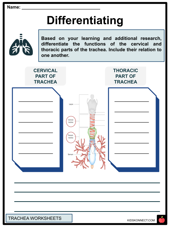
- The trachea occupies nearly the midline (slightly tipped to the left) between the larynx and the thorax. It is split into two sections:

CERVICAL PART
- The cervical segment of the trachea is located in the anterior visceral (pretracheal) section of the neck. It starts at the level of vertebra C6 at the cricoid cartilage, which forms the inferior border of the larynx and ends at the level of the superior mediastinum’s top edge and the sternum’s jugular notch.
THORACIC PART
- The superior mediastinum of the thorax contains the thoracic portion of the trachea. It starts at the outstanding thoracic opening and ends at the bifurcation of the trachea. It bifurcates anywhere between the levels of the seventh thoracic and fourth vertebrae. It is usually situated between vertebra T5 and the sternal angle.
- The tracheobronchial tree comprises the trachea and bronchi. The thoracic region splits into the right and left primary bronchi in the tracheal bifurcation. The carina, a cartilaginous ridge that faces sagittally, is located at the tracheal bifurcation.
- Compared to the left primary bronchus, the right one is shorter, broader, and nearly vertical in its journey. It is hence more vulnerable to blockages caused by foreign bodies. To supply the lungs, each main bronchus splits into progressively smaller intrapulmonary bronchi:
- Lobar bronchi
- Segmental bronchi
- Intrasegmental (subsegmental) bronchi
- Lobar bronchi move air in the direction of the pulmonary lobes. The right main bronchus splits into three lobar bronchi for the three lobes of the right lung. The two lobar bronchi (superior and inferior) for the two lobes of the left lung are formed by the division of the left main bronchi.
- Segmental bronchi aerate the bronchopulmonary segments. The three lobar bronchi on the right side separate into ten to twelve segmental bronchi in total, one for each bronchopulmonary component. Like the right, the two lobar bronchi on the left branch are into eight to ten segmental bronchi.
- Intrasegmental (subsegmental) bronchi carry air further into the bronchopulmonary regions. Each segmental bronchus has about fifteen intrasegmental bronchi. The intrasegmental bronchi divide into many bronchioles, which give rise to the pulmonary lobules and alveoli.
BLOOD SUPPLY AND INNERVATION THROUGH THE TRACHEA
- Tracheal branches of the inferior thyroid arteries, originating from the thyrocervical trunk, supply the trachea with arterial blood. The inferior thyroid venous plexus receives venous blood drainage and discharges it into the brachiocephalic veins.
- The pre-tracheal and paratracheal lymph nodes (cervical and thoracic) flow into the deep cervical lymph nodes.
- The pulmonary plexus provides innervation to the trachea. The parasympathetic supply comes from the vagus nerve’s recurrent laryngeal nerves, which are branches.
FUNCTION
- The trachea is a part of the respiratory system’s conducting zone. Its primary job is to move oxygenated air from the upper respiratory tract to the alveoli, where it will be exchanged for gas.
- The trachea additionally defends the respiratory system through physiological and immunological means. Besides oxygen and carbon dioxide, environmental air contains several potentially dangerous substances, including germs, trash, gases, and chemicals.
- To help you breathe, the trachea collaborates with the rest of your respiratory system. Inhaling causes air to move:
- Entering your trachea from your lips and nose
- Into your left and right bronchi from your trachea
- Via the bronchi of your lungs and into the bronchioles
- The human body converts carbon dioxide from the air you breathe in tiny sacs in your lungs called alveoli (gas exchange)
- Your body makes these movements backward as you exhale to remove carbon dioxide from your body.
CONDITIONS AND DISORDERS THAT AFFECT THE TRACHEA
- Tracheal cancer can develop from cancer in other organs migrating to other areas of the body, or it can start in the trachea.
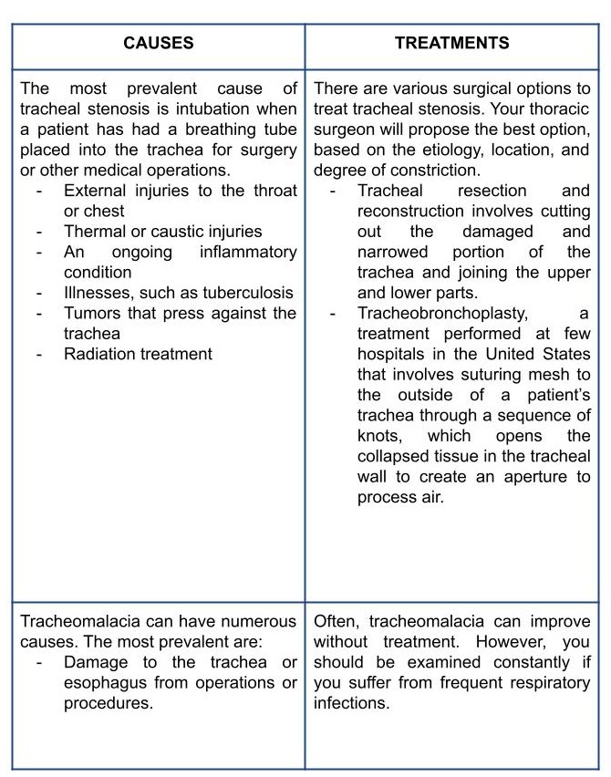
- Tracheal stenosis occurs when your trachea develops inflammation or scar tissue, which narrows it and makes breathing more challenging. Tracheal stenosis can be acquired or congenitally present.
- A tracheoesophageal fistula (TEF) is an improper connection between your esophagus and trachea. Congenital means that the condition developed while the fetus was still growing. TEF can also develop as an adult due to malignancy, an infection, or trauma.
- Tracheomalacia is an illness that mainly affects babies. It occurs when the trachea’s cartilage hasn’t properly grown. The signs of tracheomalacia include persistent respiratory infections, loud breathing, and frequent coughing.
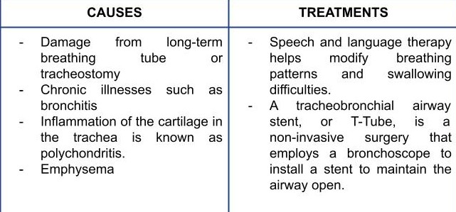
CAUSES AND TREATMENTS OF TRACHEAL DISORDERS
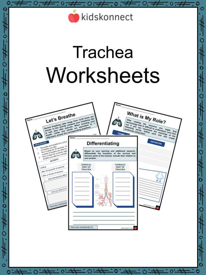
Trachea Worksheets
This fantastic bundle includes everything you need to know about the Trachea across 27 in-depth pages. These ready-to-use worksheets are perfect for teaching kids about the Trachea. The trachea, often known as the windpipe, is a respiratory organ with a diameter of less than an inch and a length of around 4 inches. The trachea allows the passage of air from the larynx to the lungs.
Complete List of Included Worksheets
Below is a list of all the worksheets included in this document.
- Trachea Facts
- The Trachea
- Source Analysis
- Differentiating
- What is My Role?
- Let’s Breathe
- Be Clean
- What is that Riddle?
- Trace it!
- Prevention
- Trachea Slogan
Frequently Asked Questions
What is the trachea?
The trachea, also known as the windpipe, is a tube-like structure made of cartilage rings and connective tissue. It extends from the larynx (voice box) to the bronchi, which are the airways that lead to the lungs.
What is the function of the trachea?
The trachea’s main function is to allow air to flow in and out of the lungs. It is a vital part of the respiratory system, which enables us to breathe and exchange oxygen and carbon dioxide.
How does the trachea stay open?
The trachea is kept open by the rings of cartilage that encircle it. These rings are incomplete at the back, allowing the trachea to expand slightly when we inhale. The trachealis muscle, a smooth muscle layer at the back of the trachea, contracts during coughing to force out any trapped material.
What are some common conditions that can affect the trachea?
Some common conditions that can affect the trachea include inflammation (tracheitis), infection (tracheobronchitis or croup), obstruction (from a foreign object, tumor, or swollen lymph nodes), and narrowing or collapse (from trauma, prolonged intubation, or certain medical conditions).
How can tracheal conditions be treated?
Treatment for tracheal conditions depends on the underlying cause. Mild cases of inflammation or infection may be treated with rest, fluids, and medications to relieve symptoms. Obstruction or narrowing may require more aggressive treatment, such as removal of the foreign object or surgery to repair the trachea. In some cases, a tracheostomy (surgical opening in the trachea) may be necessary to bypass the obstruction.
Link/cite this page
If you reference any of the content on this page on your own website, please use the code below to cite this page as the original source.
Link will appear as Trachea Facts & Worksheets: https://kidskonnect.com - KidsKonnect, May 4, 2023
Use With Any Curriculum
These worksheets have been specifically designed for use with any international curriculum. You can use these worksheets as-is, or edit them using Google Slides to make them more specific to your own student ability levels and curriculum standards.