Premium  Download Edit
Download the Anatomy Facts & Worksheets
Click the button below to get instant access to these worksheets for use in the classroom or at a home.
Download This Worksheet
This download is exclusively for KidsKonnect Premium members!
To download this worksheet, click the button below to signup (it only takes a minute) and you'll be brought right back to this page to start the download!
Sign Me Up
Edit This Worksheet
Editing resources is available exclusively for KidsKonnect Premium members.
To edit this worksheet, click the button below to signup (it only takes a minute) and you'll be brought right back to this page to start editing!
Sign Up
Not ready to purchase a subscription? Click to download the free sample version Download sample
Download This Sample
This sample is exclusively for KidsKonnect members!
To download this worksheet, click the button below to signup for free (it only takes a minute) and you'll be brought right back to this page to start the download!
Sign Me Up
Table of Contents
The study of the body’s structure is called anatomy. It is a branch of science that investigates the structures, organs, cells, and bones of animals and humans.
See the fact file below for more information on Anatomy, or you can download our 33-page Anatomy worksheet pack to utilize within the classroom or home environment.
Key Facts & Information
HISTORY OF HUMAN ANATOMY
- In 1600 BCE., the ancient Egyptians had already worked on the heart and blood vessels, especially during mummification.
- Hippocrates, a Greek physician who lived around 400 BCE, became known as the “father of medicine” and the “founder of anatomy.”
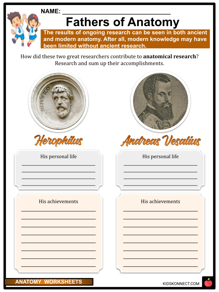
- Herophilus, a Greek who spent much of his life working in Egypt, became known as the “father of anatomy” and was one of the first to dissect human bodies.
- In Alexandria, Egypt, in 300 BCE, a school of anatomy was established.
- The most prominent physician in Ancient Greece was Claudius Galen (129-199). His conclusions are solely based on the study of animals, and his theories about human anatomy dominated and influenced medical science for more than 1,000 years.
- Soon, Herophilus and Erasistratus were the first to carry out systematic human dissections and were responsible for many discoveries made there.
- Even though the Church does not explicitly prohibit anatomy, social authorities did not allow the dissection of human remains until the 12th and even 13th centuries.
- Anatomical research stagnates as a result. Only in the 13th and 14th centuries did attitudes toward anatomy education shift. However, teaching primarily consists of lectures based on Galen’s canonical works without actual dissections to verify them.
EARLY ANATOMISTS
- It was believed that Alessandra Giliani, an Italian natural historian, who lived from 1307 to 1326 is known as the first woman to be mentioned in historical documents as practicing anatomy and pathology.
- The first accurate depiction of the human spine was created by Leonardo Da Vinci, and his notes regarding his dissection of the Florentine centenarian include the first known description of liver cirrhosis and arteriosclerosis. He was the first to develop anatomy drawing techniques that used cross-sections and multiple angles to convey information.
- “De Humani Corporis Fabrica,” written by Andreas Vesalius, was the first work to challenge Galen’s anatomical teachings and argue that they are based on observations of other mammals rather than human bodies. The anatomical components of the human body were illustrated in detail in the book.
- In order to obtain permission to dissect victims from the gallows without fear of persecution, Vesalius traveled all the way from Leuven to Padua.
- Anatomy became a real discipline under Vesalius. He made a public demonstration in 1540 of the errors in Galen’s anatomical theories, which are still accepted medical wisdom.
- Physicians and the general public soon took an interest to see the human body in person. The Greek expression “to see with one’s own eyes” is the origin of the word “autopsy.”
- Numerous cities have built anatomical theaters. People from all walks of life would attend the public dissection presentations.
- Honoré Fragonard transformed his anatomical specimens into works of art. He injected colored wax into them, and the wax stuck to the blood vessels. Varnish was applied to the remaining tissues after they dried out. His works are on display at the Ecole Nationale Vétérinaire d’Alfort in Paris.
MODERN ANATOMY
- The subdivision of anatomy that resulted from the increasingly in-depth study of operative techniques during the 18th and 19th centuries placed significant emphasis on topographic anatomy.
- Giovan Battista Morgagni pioneered the anatomical-clinical study of cadavers as a more reliable method for examining disease-related changes.
- Rudolf Virchow‘s groundbreaking discoveries in cell pathology and Pasteur and Koch’s discoveries of infectious disease agents were made possible by the emergence of pathological anatomy.
- Submicroscopic anatomy has also emerged. When applied to the study of cells, physiology, biochemistry, electron and positronic microscopy, and X-ray diffraction techniques describe their intimate structures at the molecular level.
- Imaging methods like radiography, endoscopy, angiography, computed axial tomography and tomography by positron emission, nuclear magnetic resonance imaging, echography, thermography, and others make it possible to study anatomy even in living people.
TYPES OF ANATOMY
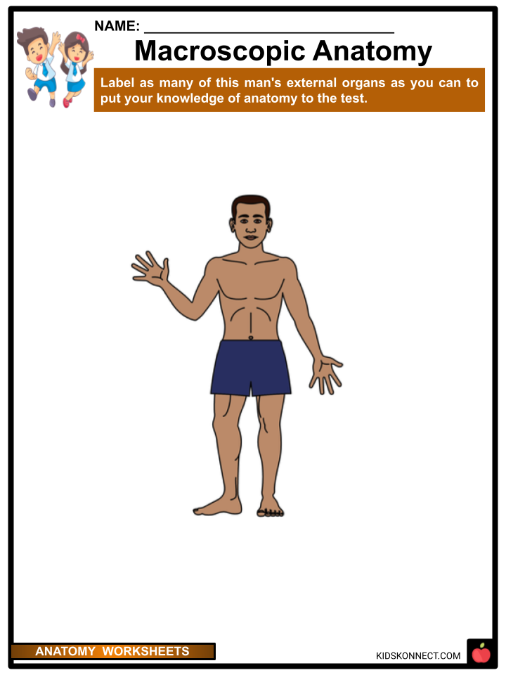
- There are two types of anatomy: Macroscopic or gross anatomy and microscopic anatomy.
- The study of anatomical features that can be seen by the naked eye is called macroscopic anatomy. It includes things like features on the outside and organs inside.
- It is further subdivided into surface anatomy (the external body), systemic anatomy (specific organ systems), and regional anatomy (specific regions of the body).
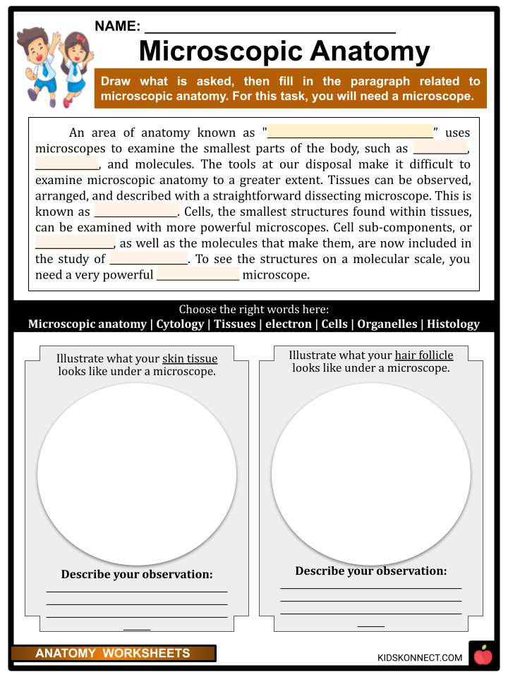
- The study of the microscopic components that make up larger structures at the level of tissues or cells is known as microscopic anatomy. Therefore, microscopes are necessary for studying microscopic anatomy.
- Histology (tissue) and cytology (cells) are additional subfields.
- Histology – The study of the microscopic anatomy of biological tissues. It looks for connections between function and structure. It aids in determining whether any treatments have improved certain diseases and their causes.
- Cytology – Studies focus on examining particular cell types, typically from fluid specimens. It is mostly used to screen for and diagnose cancer, particularly cervical cancer (pap smear), fetal abnormalities, and certain infectious agents.
- However, there are additional subdivisions of anatomy that are more specific. These include radiographic anatomy, pathological anatomy, embryology, and developmental anatomy.
- Embryology is the study of the structures that emerge between the fertilized egg and the eighth week in utero. It also investigates the production and fertilization of gametes (sex cells) as well as congenital disorders that occur before birth (teratology).
- Because it is concerned with the development of structures from the fertilized egg to the adult form, developmental anatomy studies a longer time frame.
- Radiographic anatomy is the study of body structures that can be evaluated using x-rays.
- Pathological anatomy investigates the macroscopic and microscopic changes caused by diseases.
RELATION OF ANATOMY AND PHYSIOLOGY
- Physiology is the study of how those structures work, while anatomy is about the body’s internal and external structures and their physical relationships.
- Anatomy teaches us how the body is structured, how it is held together, and how different parts interact with one another. Physiology, on the other hand, explains why these parts function in the first place.
IMPORTANCE OF UNDERSTANDING HUMAN ANATOMY
- Anatomy and physiology play a crucial role in our lives by describing our physical form and how we interact with our physical environment.
- Classes in anatomy and physiology teach us everything we need to know about our bodies, but most of us never stop to think about why they are so important.
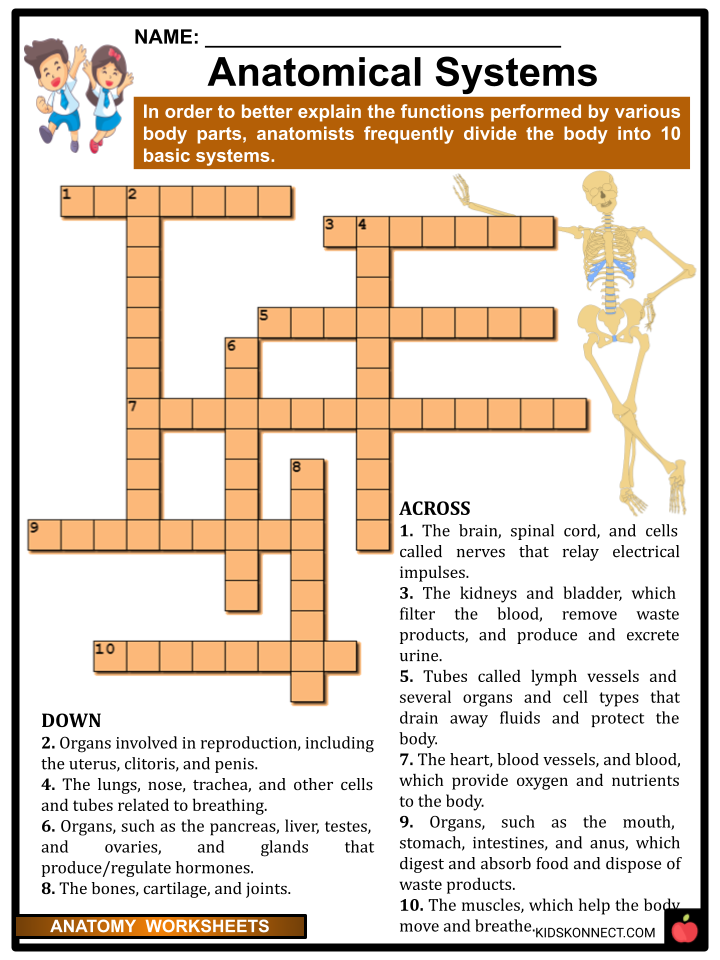
- Learning about the eleven organ systems: Digestive System, Integumentary System, Cardiovascular System, Skeletal System, Muscular System, Nervous System, Endocrine System, Lymphatic System, Respiratory System, Urinary System, and Reproductive System enables us to understand their functions and the care we need to take.
THE ORGAN SYSTEMS IN BRIEF
- Digestive System – The digestive system consists of the digestive tract and other organs that aid the body in the breakdown and absorption of food. It is a long, curled tube that begins in the mouth and travels through the esophagus, stomach, small intestine, large intestine, and towards the anus.
- Integumentary System – The integumentary system is the body’s largest organ, forming a physical barrier between the external and internal environments it protects and maintains. The epidermis, dermis, hypodermis, associated glands, nails, and hair make up the integumentary system.
- Cardiovascular System – The cardiovascular system is also known as the blood-vascular system or the circulatory system. It is made up of the heart, a muscular pumping device, and a closed system of vessels known as veins, arteries, and capillaries.
- Skeletal System – The skeletal system is the central framework of the human body. It is made up of bones and connective tissue, such as tendons, cartilage, and ligaments. It is also known as the musculoskeletal system.
- Muscular System – It is composed of specialized cells called muscle fibers. Their major function is contractibility. Movement is controlled by muscles, which are attached to bones or internal organs and blood vessels. Muscle contraction causes nearly all movement in the body.
- Nervous System – The nervous system is divided into two parts: the central nervous system (the brain and spinal cord) and the peripheral nervous system (nerves that branch off from the spinal cord and extend to all parts of the body).
- Endocrine System – Organs and glands make up the endocrine system, which is a complex network. It controls and coordinates the human body’s metabolism, reproduction, growth and development, energy level, injury response, stress, and mood hormones.
- Lymphatic System – The lymphatic system is a delicate tube network that runs throughout the body. It drains lymph fluid that has leaked from the blood vessels into the tissues and returns it to the bloodstream via the lymph nodes. The lymphatic system’s main roles include fluid management in the body.
- Respiratory System – The respiratory system is a network of organs and tissues that aid breathing. Your lungs, blood vessels, and airways are all a part of it. The respiratory system includes the muscles that power your lungs. These components collaborate to transport oxygen throughout the body and eliminate waste gases such as carbon dioxide.
- Urinary System – The function of the urinary system is to filter blood and produce urine as a byproduct. The bladder, kidneys, ureters, renal pelvis, and urethra are the organs of the urinary system. The body converts nutrients from food into energy.
- Reproductive System – The tissues, glands, and organs involved in reproduction. It has functions: to produce egg and sperm cells – which must be transported, sustained, and nurtured while developing.
- Male – prostate, testes, and penis
- Female – ovaries, fallopian tubes, uterus, cervix, and vagina.
Anatomy Worksheets
This fantastic bundle includes everything you need to know about Anatomy across 33 in-depth pages. These ready-to-use worksheets are perfect for teaching kids about Anatomy. The study of the body’s structure is called anatomy. It is a branch of science that investigates the structures, organs, cells, and bones of animals and humans.

Complete List of Included Worksheets
Below is a list of all the worksheets included in this document.
- Anatomy Facts
- Fathers of Anatomy
- Macroscopic Anatomy
- Anatomical Systems
- Anatomical Processes
- Under the Ribs
- Largely Anatomical
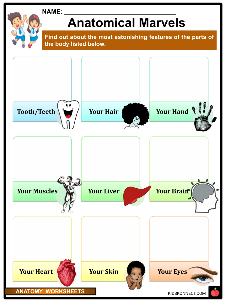
- Anatomical Marvels
- Microscopic Anatomy
- Anatomy and Physiology
- Work as One
- A Beautiful Body
- Taking Care of My Body
Frequently Asked Questions
What is the function of the pancreas in the human body?
The pancreas is an organ located behind the stomach that plays a vital role in digestion and blood sugar regulation. It produces digestive enzymes that break down carbohydrates, proteins, and fats in food, and it also produces hormones such as insulin and glucagon that regulate blood sugar levels.
What is the difference between the somatic and autonomic nervous systems?
The somatic nervous system controls voluntary movements and transmits sensory information to the brain, while the autonomic nervous system controls involuntary functions such as heart rate, breathing, and digestion. The autonomic nervous system is further divided into the sympathetic and parasympathetic branches, which have opposing effects on bodily functions.
What are the four main types of tissue in the human body?
The four main types of tissue in the human body are epithelial, connective, muscle, and nervous tissue. Epithelial tissue covers the body’s surfaces and lines internal organs, connective tissue supports and connects different structures in the body, muscle tissue allows for movement, and nervous tissue transmits and processes information throughout the body.
What is the role of the lymphatic system in the human body?
The lymphatic system is a network of vessels and tissues that help maintain fluid balance in the body and defend against infection. It collects excess fluid from tissues and returns it to the bloodstream, and it also filters lymph (a fluid that contains immune cells) and removes pathogens and other foreign substances.
What is the function of the liver in the human body?
The liver is a large organ located in the upper right abdomen that has many important functions. It produces bile, which helps digest fats, and it also processes and stores nutrients such as glucose, vitamins, and minerals. The liver also plays a key role in detoxification, breaking down and eliminating harmful substances from the body.
Link/cite this page
If you reference any of the content on this page on your own website, please use the code below to cite this page as the original source.
Link will appear as Anatomy Facts & Worksheets: https://kidskonnect.com - KidsKonnect, April 13, 2023
Use With Any Curriculum
These worksheets have been specifically designed for use with any international curriculum. You can use these worksheets as-is, or edit them using Google Slides to make them more specific to your own student ability levels and curriculum standards.


