Premium
 Download
Edit
Download
Edit
Download the Sternum Facts & Worksheets
Click the button below to get instant access to these worksheets for use in the classroom or at a home.
Download This Worksheet
This download is exclusively for KidsKonnect Premium members!
To download this worksheet, click the button below to signup (it only takes a minute) and you'll be brought right back to this page to start the download!
Sign Me Up
Edit This Worksheet
Editing resources is available exclusively for KidsKonnect Premium members.
To edit this worksheet, click the button below to signup (it only takes a minute) and you'll be brought right back to this page to start editing!
Sign Up
Not ready to purchase a subscription? Click to download the free sample version Download sample
Download This Sample
This sample is exclusively for KidsKonnect members!
To download this worksheet, click the button below to signup for free (it only takes a minute) and you'll be brought right back to this page to start the download!
Sign Me Up
Table of Contents
The sternum, also known as the breastbone, is a long, flat bone in the chest that protects the underlying muscles, organs, and major arteries, including the lungs and heart. It also serves as a link between the upper ribs on either side of the body.
See the fact file below for more information on the Sternum, or you can download our 28-page Sternum worksheet pack to utilize within the classroom or home environment.
Key Facts & Information
BACKGROUND
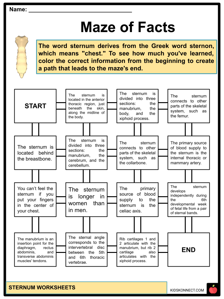
- The word sternum is derived from the Greek word sternon, which means “chest.”
- The sternum is located in the anterior thoracic region, beneath the skin, along the body’s midline. You can feel it if you put your fingers in the center of your chest.
- The sternum is a flat bone about six inches long, an inch wide, and only a fraction of an inch thick. It is one of the body’s most prominent and longest flat bones, resembling a necktie. The sternum is longer in men than in women.
- The sternum is divided into three sections: the manubrium, the body, and the xiphoid process.
STRUCTURE
- The sternum is angled obliquely, downward, and forward in its natural position. It is slightly convex in front and concave at the back; broad above, shaped like a “T”, narrowing at the point where the manubrium joins the body, then widening again to below the middle of the body and narrowing to its lower extremity.
- In children, cartilage connects the three sections of the sternum. During adulthood, the cartilage ossifies into bone.
Manubrium
- Latin for “handle,” it is the uppermost part of the sternum. It has a quadrangular shape that narrows from the top, giving it four borders. It connects to the body of the sternum, the clavicles, and the first pair of rib cartilages.
- The superior aspect of the manubrium is concave, resulting in the jugular notch, which is visible beneath the skin.
- The inferior border is oval and rough, with a thin layer of cartilage covering it for articulation with the body.
Body
- It is the longest part of the sternum. It has flat sides with depressed ridges where the costal cartilages of the third to seventh pairs of ribs articulate inferior to the sternal angle.
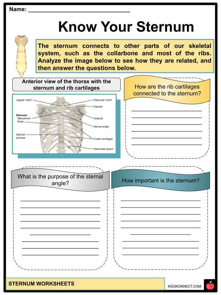
- The sternal angle is formed by the junction of the body and the manubrium.
- It articulates superiorly with manubrium and inferiorly with the xiphoid process.
Xiphoid Process
- It is the most minor and inferior part of the sternum. Its shape and size vary, with its tip located at the level of the T10 vertebrae. The xiphoid process has a cartilaginous structure and ossifies later in life, around 40.
- The xiphoid process articulates with a part of the costal cartilage of the seventh rib in some people.
FUNCTION
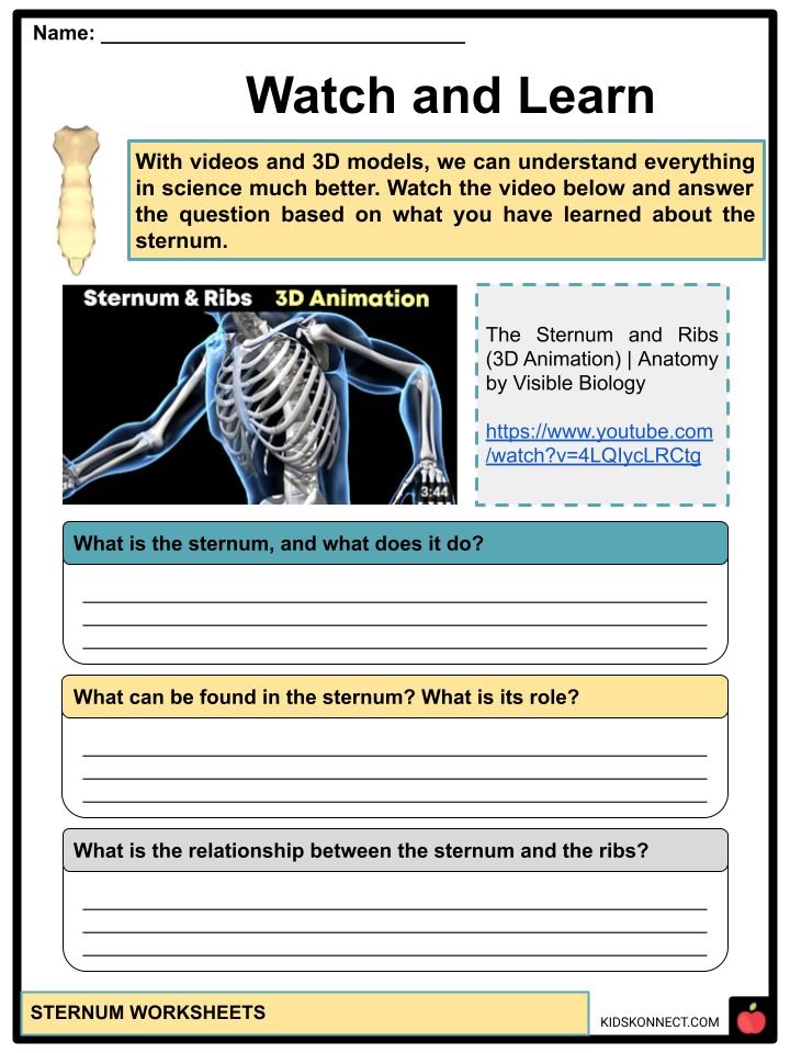
- The primary function of the sternum is to protect the underlying organs from injury. The sternum, along with the ribs, protects the torso organs such as the heart, lungs, and chest blood vessels.
- The sternum also connects to other parts of the skeletal system, such as the collarbone and most of the ribs.
- The sternum is also connected to some muscles in the chest and upper abdomen.
- The xiphoid process is an insertion point for the diaphragm, rectus abdominis, and transverse abdominis muscle tendons.
- The sternal angle corresponds anteriorly to the intervertebral disc between the 4th and 5th thoracic vertebrae. This angle is also known as the Louis sternal angle.
ARTICULATION
- Rib cartilages 1 and 2 articulate with the manubrium, but rib two cartilage also articulates with the body of the sternum.
- The primary articulations with the body of the sternum are cartilages 2, 3, 4, 5, 6, and 7.
- The sternal angle creates the “bucket handle,” which moves up and outward, with the superior end of the manubrium remaining relatively fixed.
- The sternum is also attached to several muscles (neck, thorax, and anterior abdominal wall muscles). Attached to the manubrium are the sternocleidomastoid, sternohyoid, and sternothyroid. The transversus thoracis muscle connects to the body and the xiphoid process.
- The primary source of blood supply to the sternum is the internal thoracic or mammary artery (ITA). It descends 1-2 cm from the lateral margin of the sternum to the posterior aspect of the chest wall.
DEVELOPMENT
- The sternum develops independently during the 6th developmental week of fetal life from a pair of sternal bands called “sternal bars.”
- These two sternal bands emerge bilaterally by the 10th week of intrauterine life, then convert to pre-cartilaginous structures to form the sternal plate.
- The mesenchyme condenses in the 7th week of intrauterine life, forming a primary cartilaginous model of the three sternal segments (manubrium, body, and the xiphoid process).
- The cartilaginous sternal model is made up of six horizontal divisions called sternebrae. The manubrium and xiphoid process is represented by the superior and inferior sternebrae. The body of the sternum is represented by the four sternebrae that lie between them. The manubrium is the first part of the sternum to form during embryogenesis, followed by the sternal body and the xiphoid process.
SURGICAL CONSIDERATIONS
Sternotomy
- Sternotomy is considered the standard gold incision for cardiac surgery. The median sternotomy is a common critical incision in which the surgeon splits the sternum along the median plane, allowing the surgeon to see the heart, great vessels, and lungs better. It is the most common type of osteotomy performed worldwide.
Sternal Aspiration and Sternal Biopsy
- These are procedures used to collect bone marrow specimens from the sternum. The sternum is a common site for bone marrow collection because sternal hematopoietic marrow persists throughout life. Bone marrow examination is frequently indicated for diagnosing blood dyscrasias and metastatic cancer.
MALFUNCTIONS AND DEFECTS
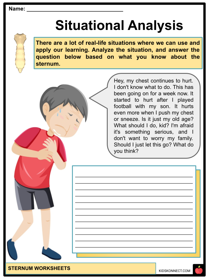
Fracture of the sternum
- A rare fracture that is most commonly caused by high-impact direct trauma. The manubrium is the most commonly injured bone part. The ribs frequently fracture during the process due to their direct connection and proximity. The vital organs may be jeopardized.
Pectus excavatum
- Also called funnel chest, it is distinguished by the inward displacement of the adjacent costal cartilages of the sternum, resulting in an abnormal depression of the anterior chest wall.
Pectus Carinatum
- This condition occurs when the ribs and sternum grow abnormally, causing the sternum to protrude outwards.
Sternal cleft
- An extremely rare congenital abnormality caused by sternum fusion failure, this condition results in a sternal cleft, which can be seen at birth without any symptoms.
Sternal foramen
- A relatively uncommon congenital sternum disorder is sometimes referred to as an anatomical variation. It is a single round hole in the sternum that is present from birth and is usually off-centered to the right or left, commonly forming in the second, third, and fourth segments of the breastbone body.
- Congenital sternal foramina are frequently mistaken for bullet holes. They are usually asymptomatic but can be problematic if acupuncture is intended for the area.
ANIMALS
- In vertebrate anatomy, the sternum is a flat bone located in the middle front part of the rib cage. It is endochondral in origin.
- It most likely evolved as an extension of the pectoral girdle in early tetrapods; it is not found in fish. It is usually a shield-shaped structure made entirely of cartilage in amphibians and reptiles.
- It is not found in turtles or snakes. In birds, the sternum is a relatively large bone with an enormous projecting keel to which the flight muscles are attached. Only mammals have the segmented, elongated sternum that is seen in humans.
Sternum Worksheets
This is a fantastic bundle that includes everything you need to know about the Sternum across 28 in-depth pages. These are ready-to-use worksheets that are perfect for teaching kids about the Sternum, which is a long, flat bone in the chest that protects the underlying muscles, organs, and major arteries, including the lungs and heart.

Complete List of Included Worksheets
Below is a list of all the worksheets included in this document.
- Sternum Facts
- Sternum Sketch
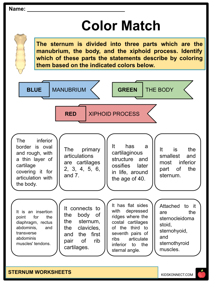
- Color Match
- Sternum Puzzle
- Know Your Sternum
- Maze of Facts
- Defects Scrabble
- Watch and Learn
- Which Animal?
- Situational Analysis
- 3-2-1
Link/cite this page
If you reference any of the content on this page on your own website, please use the code below to cite this page as the original source.
Link will appear as Sternum Facts & Worksheets: https://kidskonnect.com - KidsKonnect, November 10, 2022
Use With Any Curriculum
These worksheets have been specifically designed for use with any international curriculum. You can use these worksheets as-is, or edit them using Google Slides to make them more specific to your own student ability levels and curriculum standards.